In the current field of medical disease research, clinical patients are still the most reliable subjects for disease research, but for ethical reasons and other limitations of not being able to obtain samples directly from living human beings, there is an urgent need to develop other stable, reliable, and reproducible disease research models that can replace the human-subject-based disease models.
Taking normal or patient-derived cells or tissues as research objects, in vitro construction of models with structural and functional characteristics similar to those of human tissues and organs is an important area of disease research, in which organoid and organ chip are the most promising technologies expected to solve the above challenges.

Traditional in vitro disease modeling consists mainly of cellular models that are cultured in a predominantly two-dimensional (2D) static manner. Cells cultured in this way are able to maintain some of their biological functions, but lack the necessary microenvironmental factors such as multicellular interactions, cell-extracellular matrix (ECM), and physicochemical stimuli in vivo, which makes a single static cell culture unable to realistically mimic the physiological and pathological states of human tissues and organs.
Although animal models can simulate the physiological and pathological processes of multi-cells, multi-tissues and organs interacting with each other, limitations such as species and genetic background variability as well as animal ethics make it difficult to fully reproduce in animals a disease that occurs in humans, and may even lead to conclusions that are contrary to those of human diseases.
Organoid is a “mini” organ containing multiple cellular components with structural and functional characteristics of source tissues and organs formed by pluripotent stem cells and adult stem/precursor cells through cell self-assembly in vitro.
It gradually demonstrates its great application prospects in the fields of simulating human tissue and organ development, organism homeostasis, disease mechanism research and drug development.
At present, organoid culture methods mainly include matrix gel embedding, suspension dynamic culture means, through the addition of nutrients and specific growth factors and so on in the appropriate culture medium, to achieve the promotion of cell division and proliferation, directional differentiation, as well as the formation of spatial structure.
Ensure that organoids maintain both the self-renewal of stem cells and the maintenance of specific structures and functions by differentiated cells in a specific culture system.
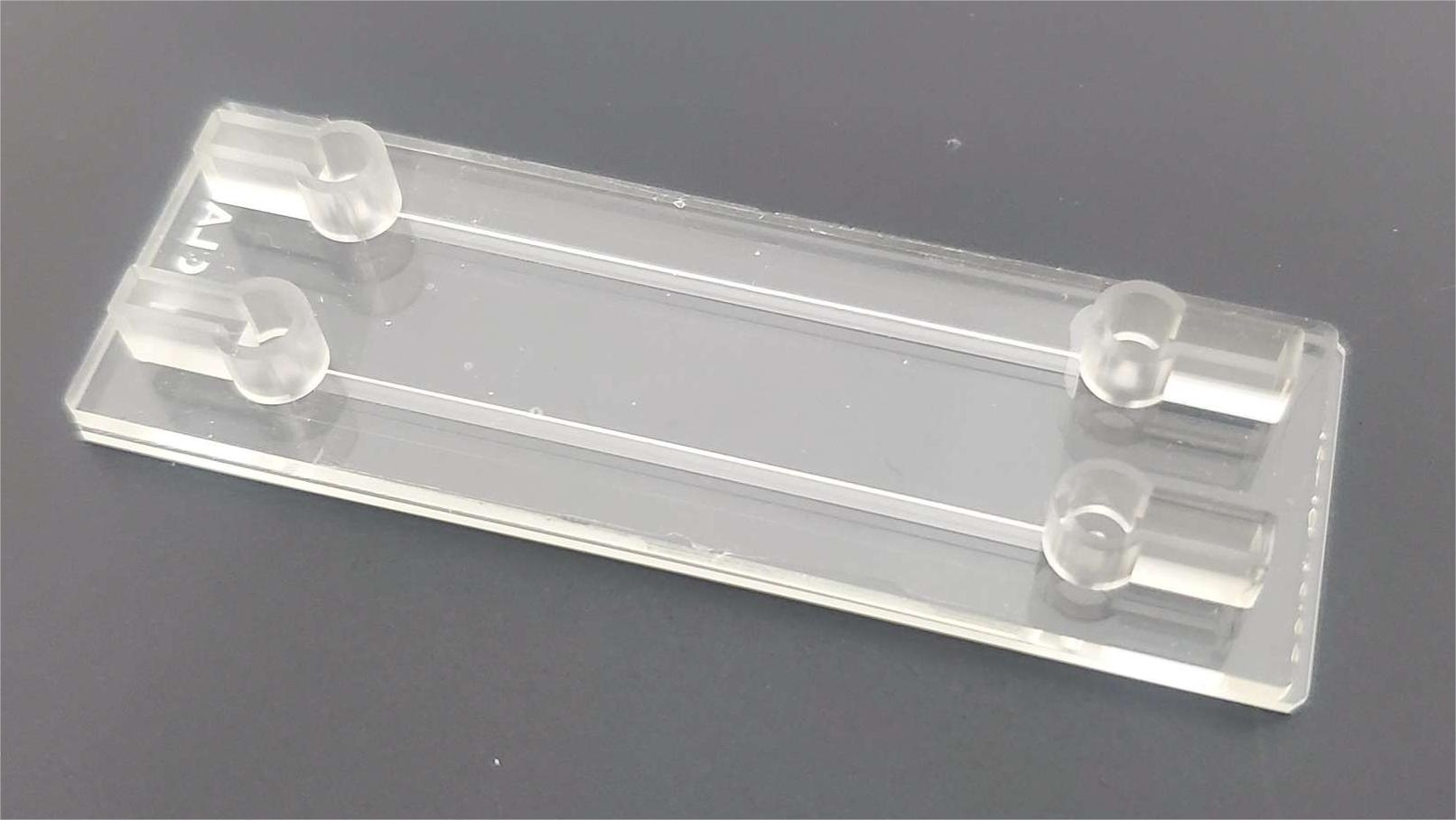
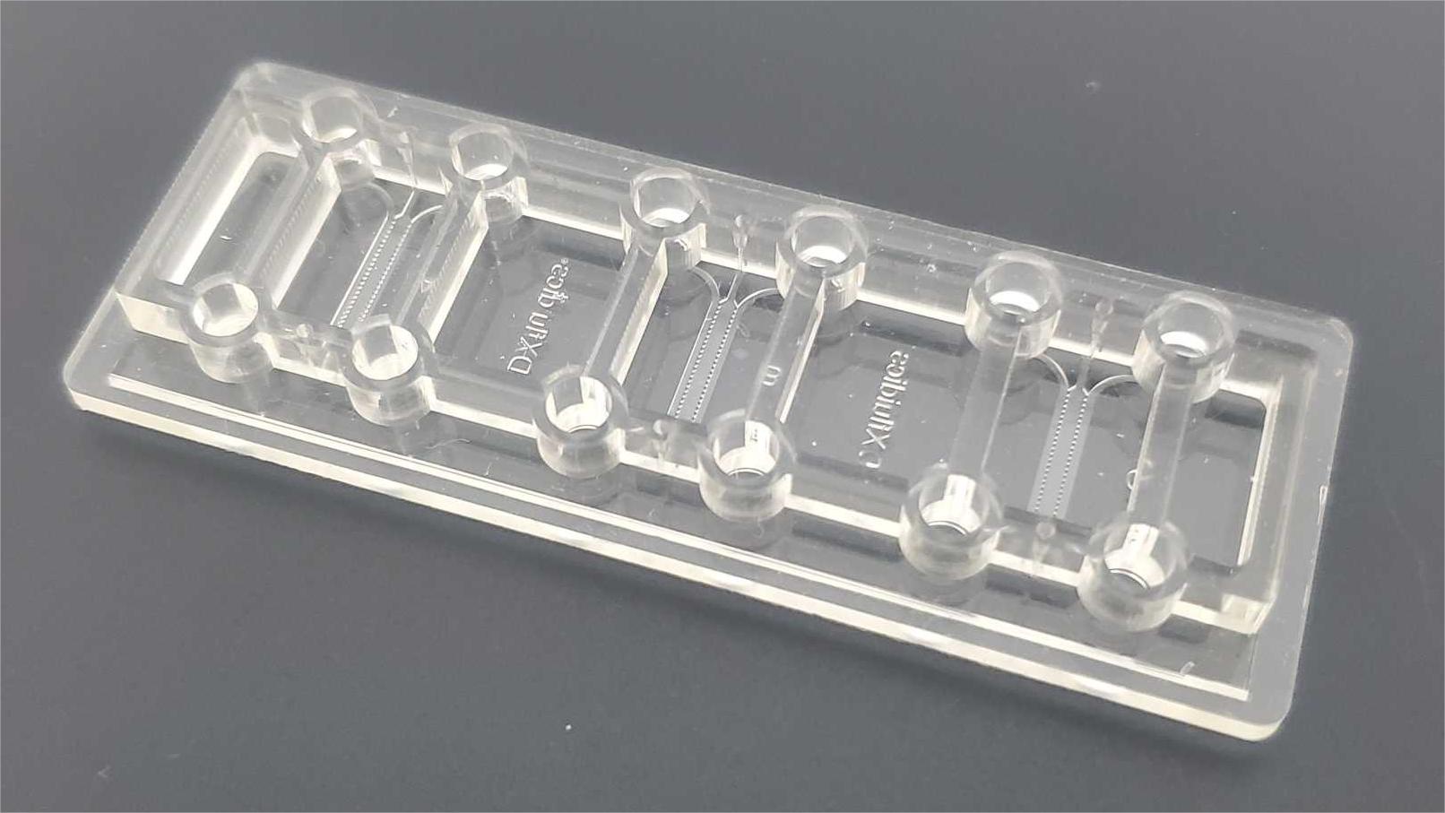
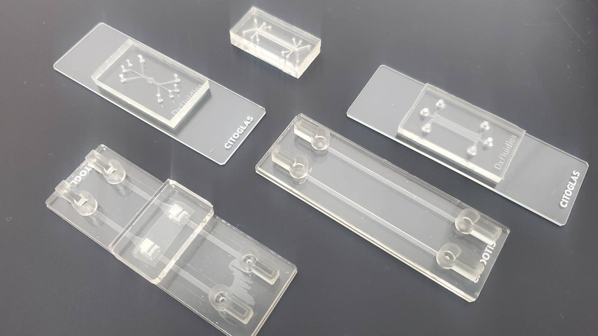
Organ-on-a-chip (OOC) is a transformative new technology that has been rapidly developed in the last decade. It is a micro cell/tissue culture carrier based on microfluidic chip technology to simulate the functional units of human tissues and organs in vitro, including the basic components and elements necessary for the functional units, such as multicellular components, extracellular matrix, and microenvironmental physical and chemical factors.
OrganoChips can make up for the shortcomings of existing cell culture methods, and have advantages that are unmatched by traditional methods: including three-dimensional dynamic culture, controlled physicochemical stimulation, low cost, high throughput, high reliability, and so on.
At the same time, organ chips combine with imaging instruments to monitor cell biological changes in real time, etc., to better record the behavioral changes of cells in disease states and the whole process of response to drugs.

Organoids-on-a-chip is a combination of the advantages of both organoid and organ chip technologies, which enables more accurate and controllable organoid model construction by integrating microfluidic technology, which can control and simulate the microenvironment, vascularization, and inter-tissue interactions of tissues, and facilitate high-throughput analysis of organoid models.
Combining the advantages of medical biology and engineering, organoid chip can effectively solve the limitations of traditional organoid culture technology, which not only realizes 3D culture of cells and reproduces the interrelationships between cells and between cells and matrix, but also simulates the important in vivo microenvironments of target organs in vivo, such as interstitial fluids and mechanical force stimulation.
Through real-time imaging, sensors and other technologies, organoid microarrays have unparalleled advantages in bionic microenvironments for the study of individual genetic development, mechanisms of disease development, drug screening and immune evaluation.
The small size of organoid chip and its multiple culture cavities can improve the experimental throughput, which is suitable for the synchronization of multi-group control experiments. It can be used in the construction of organ development model or disease model, drug development, immune evaluation and other fields, which greatly reduces the individual variability and experimental cycle, makes the research of single organoid possible, and provides a strong argumentation for preclinical research.
Key application areas for organoid chips
Construction of organ development models and research on developmental biology
Organoid microarrays can accurately mimic the tissue structure of the target organ, use microchannels as a source and distribution pathway for soluble factors, control the distribution of biochemical concentration gradients in the ECM, and induce tissue regionalization similar to that in vivo.
For example, combining microchannels with patterned culture scaffolds confines stem cells and differentiated epithelial-like organs to crypts and villi, respectively, in gut-like structures.
Cell-cell interactions are important in maintaining the stability of the internal environment and signaling, etc. Organoid microarrays use integrated culture cavities to simulate multi-organ interactions in vitro.
There are two main types of organoid chip co-culture systems: contact co-culture represented by microdroplets and micro-contact printing, and non-contact co-culture represented by hydrogels, semi-permeable membranes and narrow channels.

Construction of Disease Models and Applied Research
Disease modeling is a major challenge in cancer research, including tumor development, developmental disorders, and microbial infections. Culturing patient-derived tumor cells or induced pluripotent stem cells (iPSCs) on organoid microarrays has great potential for constructing specific disease models and enabling “personalized” treatment for patients.
The lack of biological models of host cell-microbe interactions is a major impediment to human studies of microbial pathogenesis. Organoid microarrays can integrate multifunctional structural units, which to some extent can realize the different requirements of bacteria and cells for oxygen concentration and the long-term nature of co-culture experiments between somatic cells and microorganisms.
Drug development and research
Drug development requires consideration of pharmacokinetics, toxicity, and efficiency of the delivery system, but the lack of realistically controllable clinical models makes the process expensive and lengthy.
The organoid chip is widely used in drug screening and drug analysis because of its high throughput, high integration and reproducibility, which can reduce the cost of drug development.
Simulating the structure and physiological processes of different organs in bionic microenvironments and reproducing the interactions and crosstalk between organs enables more complex and physiologically meaningful drug studies.
It has been shown that cardiac-hepatic-intestinal co-culture microarrays incorporating microvessels successfully simulate the absorption, distribution, metabolism, excretion, and toxicokinetic process (ADMET) of drugs, which can reproduce the metabolic process of drugs in vivo.
High-throughput organoid microarrays using multiple chambers and compatible delivery concentration gradient generators have been a new approach to drug discovery or “personalized” drug therapy in recent years, automating the processing of multiple combinations of drugs at different concentrations in a short period of time and improving the efficiency of drug discovery.
Organoid chips can control the size and uniformity of organoids by optimizing the size of the micropillar arrays to reduce experimental errors.
immune response
Organoids lose a portion of their immune cells, key stromal cells, and cytokines during culture, which can limit functional testing of patient-derived organoids for chemotherapy and targeted drug screening.
Studies have shown that cancer and immune cell interactions are individual and organ-specific, so this dilemma may be overcome by co-cultivating tumor-like and immune cells via organoid microarrays and by microfluidically mimicking the tumor microenvironment and capturing changes in subtle dynamics.

Issues that need to be addressed urgently
The research objectives of organoid and organoid chip fabrication are, firstly, to accurately simulate the microenvironment of human organs and the functional relationship between various functional components, so as to establish disease models, carry out drug screening and toxicity testing, and provide personalized medical treatment; secondly, to be able to use tissues or organs constructed in vitro to replace diseased or aged tissues or organs in the body in the near future, and to develop clinical applications in the field of tissue engineering and regenerative medicine. The second is to be able to use in vitro constructed tissues or organs to replace diseased or aging tissues or organs in the human body in the near future, and to develop clinical applications in tissue engineering and regenerative medicine.
However, there are still many obstacles and challenges to realize the research breakthroughs in organoid and organ-like chip technology.
For one thing, the interactions between the environment and cells and between cells are intricate and complex, and there are many interactions that have yet to be discovered or studied, and this unknown creates difficulties for the goal of precisely controlling cellular interactions, and AI techniques such as big language modeling have the potential to help with this type of challenge.
Second, self-organization of organoids is an extremely dynamic process, and maintaining controllability and predictability during its development is challenging, requiring both a deeper understanding of the biological mechanisms of self-organization, real-time dynamic monitoring, and real-time and timely intervention with engineered means of regulation and intervention.
Third, complex ethical issues are involved in the application of organoid research and the construction of platforms, including the use of stem cells, the compliance of tissue sources, and the privacy and security of patient data, etc. It is necessary to reasonably adjust the ethical standards and regulations in accordance with the technological advancement, so as to ensure the protection of the rights and interests of the individuals and the legitimacy of the research in the process of the development of the technology, and at the same time, not to impede the positive advancement of the technology.
About Us
DingXu (Suzhou) Microfluidics Technology Co., Ltd. is a high-tech enterprise dedicated to the field of microfluidics. We are committed to providing customers with comprehensive microfluidic solutions, including customized microfluidic chip development, surface modification, microfluidic chip processing equipment, and microfluidic instruments. Our team boasts extensive experience and technical expertise, continuously combining professional knowledge with innovative thinking to deliver high-quality solutions. We consistently prioritize customer-centric values, embrace self-challenges, and pursue excellence. Through professionalism, innovation, and collaboration, we aim to create greater value for our customers and contribute to a brighter future in the field of microfluidics.
© 2025. All Rights Reserved. 苏ICP备2022036544号-1


